Biological structures
Posted on 14/11/2014

Anton Le Brun
Whoop whoop: pertussis toxin
What does it look like?
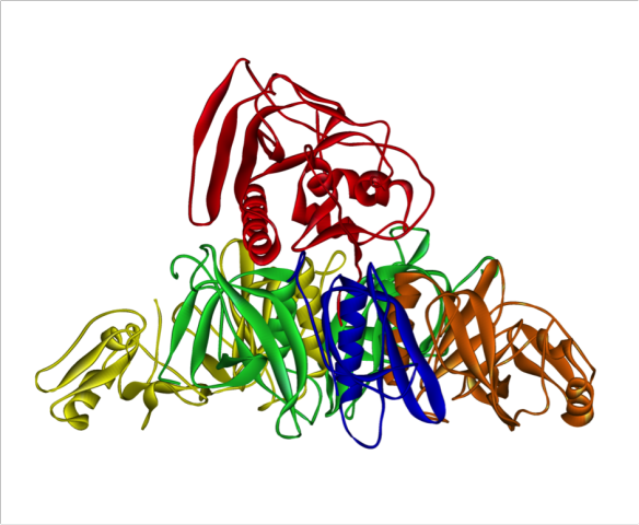
Pertussis toxin from Bordetlla pertussis.
What is it?
Pertussis toxin is released by virulent forms of the bacteria Bordetlla pertussis, the cause of whooping cough. Whooping cough gets its name from the characteristic 'whooping' sound that infected children make when coughing.
The pertussis toxin is known as an AB5 toxin as it contains an A subunit (in red), which is a catalytic subunit, and five B subunits (other colours) that transport the toxin into the cells of those infected with B. pertussis. This AB arrangement is similar to other crystal structures seen on this blog such as anthrax toxin and ricin.
The B subunits of the pertussis toxin bind to receptors on the cell membrane and the toxin is internalised into the cell. The A subunit has catalytic activity known as ADP-ribosylation, and, once inside the cell, the A subunit is activated. The A subunit modifies a protein called G-protein by bonding a molecule of ADP-ribose onto the G protein. This prevents G-protein from interacting with G-protein coupled receptors, thus disrupting the cell's internal lines of communication.
Where does it come from?
The crystal structure of pertussis toxin was first solved by Penelope Stein and colleagues at the University of Alberta, Canada in 1994 [1]. The crystal structure is from the protein data bank, PDB code 1PRT.
[1] P. E. Stein, A. Boodhoo, G. D. Armstrong, S. A. Cockle, M. H. Klein, R. J. Read: The crystal structure of pertussis toxin. Structure (1994) 2: 45-57.
Related articles
|
It takes two – Vaterite |
Fighting the Flu – The structure of Neuraminidase |
Caffeine hit: Adenosine A2A receptor |